Mammography
About Breast Cancer
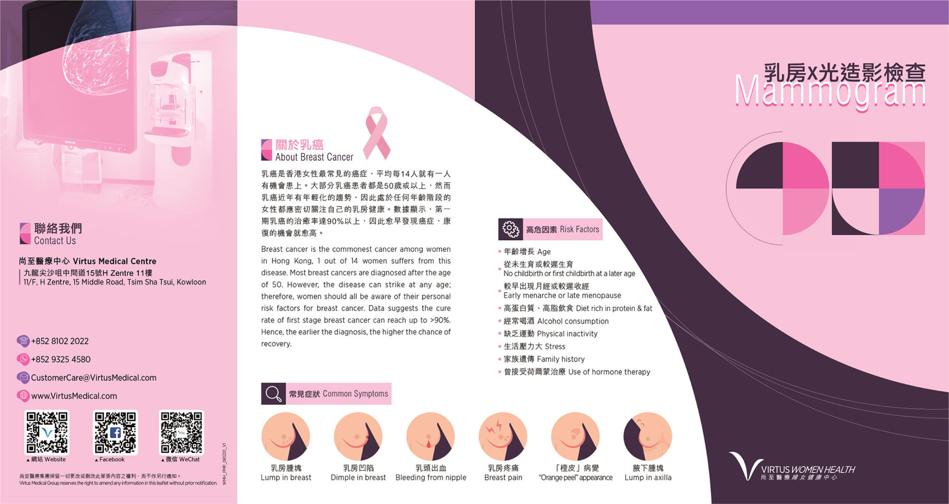
Breast cancer is the commonest cancer among women in Hong Kong, 1 out of 14 women suffers from this disease. Most breast cancers are diagnosed after the age of 50. However, the disease can strike at any age; therefore, women should all be aware of their personal risk factors for breast cancer. Data suggests the cure rate of first stage breast cancer can reach up to >90%. Hence, the earlier the diagnosis, the higher the chance of recovery.
Common Symptoms
- Lump is breast
- Dimple in breast
- Bleeding from nipple
- Breast pain
- "Orange peel" appearance
- Lump in axilla
Risk Factors
- Age
- No childbirth or first childbirth at later stage
- Early menarche or late menopause
- Diet rich in protein and fat
- Alcohol consumption
- Physical inactivity
- Stress
- Family history
- Use of hormone therapy
What is a Mammogram?
A mammogram is a low-dose X-ray examination of the breast and is one of the commonest screening and diagnostic tests for breast cancer. During a mammogram, X-ray images of each breast are taken from different angles to look for any microcalcifications, architectural distortions or masses that are too small to be felt, making mammography a useful tool for early detection of breast cancer. Additional examinations, such as breast ultrasound, magnetic resonance imaging (MRI) and biopsy, may be required for better diagnostic accuracy.
BIRADS (Breast imaging reporting and data system), a classification system developed by American College of Radiology
| Category | Definition | Likelihood of Cancer | Recommendation |
| 0 | Incomplete | NA | Further examination needed |
| 1 | Negative | 0% | Routine examination |
| 2 | Benign | 0% | Routine examination |
| 3 | Probably benign | >0% - ≤2% | Early follow-up visits or biopsy |
| 4a | Low suspicion | >2% - ≤10% | Biopsy |
| 4b | Moderate suspicion | >10% - ≤50% | Biopsy |
| 4c | High suspicion | >50% - <95% | Biopsy |
| 5 | Highly suggestive of malignancy | ≥95% | Biopsy |
| 6 | Known biopsy - proven malignancy | 100% | Cancer treatment |
2D Mammogram VS 3D Mammogram
A conventional mammogram takes 2 X-ray images of each breast, one from the front and one from the side, to create one single 2D image of the breast; and a 3D mammogram, as its name suggests, is able to produce a 3D image by combining multiple images from different angles in thin slices (around 1mm). 3D mammography provides doctors with a clearer view through the overlapping tissues and allows them to check for the slightest signs of abnormalities.
Mammogram FAQ
Is a Mammogram Painful?
No matter which type of mammogram (2D or 3D) you are having, your breast has to be compressed between two imaging plates from two different angles for X-ray images to be taken. Some may experience discomfort or even pain during the process. Luckily, 3D mammogram requires less compression and is relatively less painful.
What Is the Risk of Mammography?
The radiation risk of mammography is extremely low. The total radiation dose for a screening mammogram is about 0.36mSv. To put that number in perspective, it is equivalent to one-eighth of the total radiation dose a person absorbed annually from his/her natural surroundings, or the total radiation dose a person is exposed to while making 4 return flight journeys between Hong Kong and London. (Data source: Radiation Health Unit, Department of Health)
Who Should Get a Mammogram?
Women of age 40 or above are recommended to get a mammogram every 2 years; and for women under age 40, who are still in their childbearing year and tend to have denser breasts, breast ultrasound will be a better option. Please consult your doctor for personalized advice.
What Are the Differences between Breast Ultrasound and Mammogram?
Breast ultrasound is an examination that uses sound wave to distinguish fluid-filled cysts from solid masses. It is painless and involves no radiation. However, microcalcification typically do not show up on ultrasound. Therefore, it cannot replace mammogram and vice versa.
What Should I Do If the Result Is Suspicious?
If your result is suspicious, a biopsy will be recommended to determine whether the suspicious area is cancerous or not. After a diagnosis is made, your doctor will work with you to develop a treatment plan that meets your need.
Service Leaflets